The field of brain mapping neuroscience has revolutionized our understanding of the human brain by creating detailed spatial and functional representations of neural activity. Brain mapping neuroscience combines advanced imaging techniques, computational modeling, and experimental methods to visualize and analyze the complex networks that govern cognition, behavior, and consciousness. Since its inception in the late 20th century, brain mapping neuroscience has evolved from simple structural studies to dynamic, high-resolution mapping of neural circuits in real time. According to Dr. Karl Deisseroth, a pioneer in optogenetics and professor at Stanford University, "brain mapping neuroscience has transformed how we study neural circuits, allowing us to observe and manipulate activity with unprecedented precision" (Nature Reviews Neuroscience).
Historical Foundations of Brain Mapping Neuroscience
The origins of brain mapping neuroscience can be traced back to the early 19th century when phrenologists attempted to localize functions in the brain based on skull bumps. While phrenology was later discredited, it laid the groundwork for the idea of functional localization. In 1861, French neurologist Paul Broca identified the area responsible for speech production, now known as Broca's area, providing the first concrete evidence of functional specialization. A century later, in the 1970s, the development of computed tomography (CT) scans marked a major milestone, enabling non-invasive visualization of brain structures. The 1980s saw the advent of magnetic resonance imaging (MRI), which further refined brain mapping capabilities. By the 1990s, functional MRI (fMRI) emerged, allowing researchers to observe brain activity in real time as individuals performed cognitive tasks. According to a 2020 review in *Science*, "the evolution of brain mapping neuroscience has been driven by technological innovations that progressively enhanced spatial and temporal resolution" (Science).
Early Techniques and Limitations
In the early days of brain mapping neuroscience, researchers relied on invasive methods such as lesion studies and electrophysiology. Lesion studies involved observing behavioral changes after brain damage, while electrophysiology measured electrical activity in individual neurons. These methods provided valuable insights but were limited by their invasiveness and low spatial resolution. The introduction of PET scans in the 1970s offered a non-invasive alternative by tracking metabolic activity, but it had poor temporal resolution. The breakthrough came with fMRI in 1992, which detected blood flow changes associated with neural activity. Dr. Marcus Raichle, a neuroscientist at Washington University, noted that "fMRI revolutionized brain mapping neuroscience by enabling researchers to study the living human brain with remarkable detail" (NIH).
Modern Techniques in Brain Mapping Neuroscience
Today, brain mapping neuroscience encompasses a wide array of techniques, each offering unique advantages. High-resolution structural MRI provides detailed anatomical images, while diffusion tensor imaging (DTI) maps white matter tracts. Functional MRI captures hemodynamic responses, but its temporal resolution is limited to seconds. To overcome this limitation, researchers use electroencephalography (EEG) and magnetoencephalography (MEG), which measure electrical and magnetic activity with millisecond precision. Recent advances in optogenetics and calcium imaging allow for the manipulation and observation of neural activity in animal models with unparalleled accuracy. A 2021 study published in *Nature* demonstrated that combining fMRI with calcium imaging in mice could resolve activity at the level of individual neurons, a significant leap for brain mapping neuroscience (Nature).
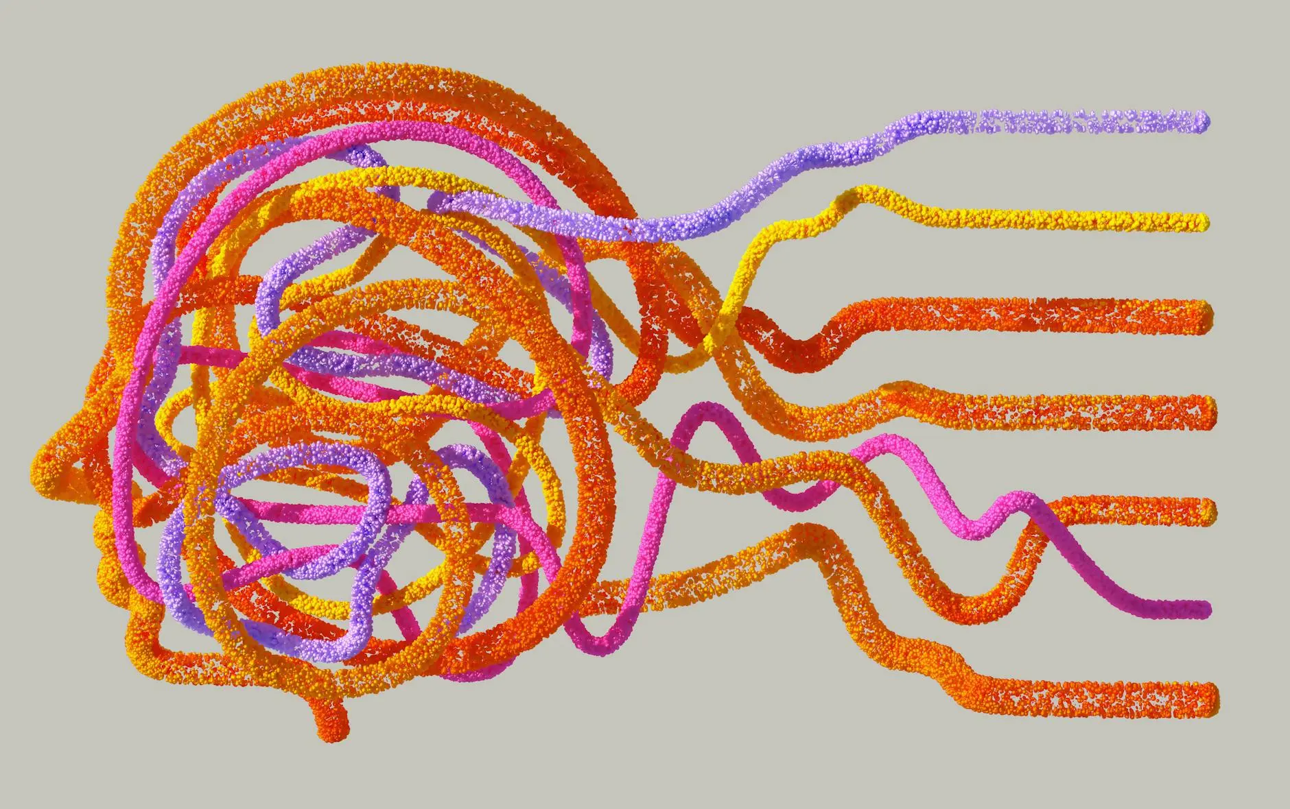
Connectomics and Network Analysis
A major focus of modern brain mapping neuroscience is connectomics—the comprehensive mapping of neural connections. The Human Connectome Project, launched in 2010, aims to create a detailed map of the brain's structural and functional connections. Using advanced MRI techniques, the project has identified over 100,000 fiber bundles and characterized their roles in cognition. Researchers reported that "the brain's functional networks exhibit both modular organization and global integration, allowing for efficient information processing" (Human Connectome Project, 2013). Machine learning algorithms are increasingly used to analyze these vast datasets, revealing patterns of connectivity associated with neurological and psychiatric disorders.
Advanced Imaging Technologies
Recent innovations in brain mapping neuroscience include ultra-high-field MRI (7T and above) and functional ultrasound imaging. These technologies offer improved spatial resolution, enabling the visualization of smaller brain structures. Functional ultrasound, which uses sound waves to detect blood flow changes, has been shown to achieve spatial resolution comparable to fMRI while being more portable. A 2022 study in *Science* highlighted the potential of functional ultrasound for clinical brain mapping, noting its "ability to monitor brain activity in real time with high sensitivity" (Science). Additionally, advances in molecular imaging allow researchers to track specific neural populations using fluorescent markers, providing insights into the molecular basis of brain function.
Applications of Brain Mapping Neuroscience
The applications of brain mapping neuroscience span basic research, clinical medicine, and technology development. In basic research, brain mapping has elucidated the neural mechanisms underlying perception, memory, and decision-making. Clinically, it aids in the diagnosis and treatment of neurological disorders such as epilepsy, Alzheimer's disease, and stroke. For example, epilepsy patients often undergo pre-surgical brain mapping to identify seizure foci while preserving critical functions like language and motor control. According to Dr. Itzhak Fried, a neurosurgeon at UCLA, "brain mapping neuroscience has transformed epilepsy surgery by allowing us to pinpoint the exact location of seizures with minimal risk to healthy tissue" (Nature).
Neurological Disorders and Mental Health
Brain mapping neuroscience has been instrumental in understanding the neural basis of mental health disorders. Studies using fMRI have identified abnormal connectivity patterns in patients with depression, schizophrenia, and autism. For instance, a 2019 meta-analysis in *Nature Neuroscience* found that "default mode network hyperconnectivity is a consistent biomarker in major depressive disorder" (Nature Neuroscience). These findings have led to the development of targeted neuromodulation therapies, such as transcranial magnetic stimulation (TMS) and deep brain stimulation (DBS), which normalize abnormal activity patterns. Brain mapping neuroscience also plays a role in early detection of Alzheimer's disease, where changes in hippocampal volume and functional connectivity can precede clinical symptoms by years.
Brain-Computer Interfaces
One of the most exciting applications of brain mapping neuroscience is the development of brain-computer interfaces (BCIs). BCIs translate neural activity into commands for external devices, enabling paralyzed individuals to control prosthetic limbs or communicate. A landmark study in 2022 demonstrated a high-bandwidth BCI that allowed a tetraplegic patient to type at 90 words per minute using neural signals decoded by brain mapping algorithms. Researchers reported that "this technology represents a paradigm shift in assistive devices, directly harnessing the brain's computational power" (Science). Future advancements in brain mapping neuroscience could lead to bidirectional BCIs, which not only read neural signals but also stimulate the brain to restore lost functions.
Challenges and Ethical Considerations
Despite its progress, brain mapping neuroscience faces significant challenges. The brain's complexity, with its billions of neurons and trillions of synapses, makes comprehensive mapping a daunting task. Current techniques often provide only partial snapshots of brain activity, missing dynamic interactions across multiple scales. Additionally, the interpretation of brain mapping data is complicated by individual variability and the brain's plasticity. Ethical concerns also arise, particularly regarding privacy and consent. As brain mapping techniques become more advanced, the potential for "mind reading" or manipulation raises questions about neuroethics. A 2021 report by the *NIH* emphasized the need for "clear guidelines on the responsible use of brain mapping data to protect individual rights and ensure equitable access" (NIH).
Data Privacy and Security
The vast amounts of data generated by brain mapping neuroscience pose significant privacy risks. Unlike other forms of medical data, brain data can reveal intimate details about thoughts, emotions, and intentions. A 2020 study in *Nature* highlighted the vulnerability of brain data to hacking, noting that "malicious actors could potentially exploit neural data to extract sensitive information or manipulate behavior" (Nature). To address these concerns, researchers are developing encryption methods and anonymization techniques. However, the ethical debate continues, with calls for stricter regulations on the collection and use of brain mapping data.
Equity and Access
Another challenge in brain mapping neuroscience is ensuring equitable access to its benefits. Advanced imaging technologies are expensive and often limited to research institutions in high-income countries. This disparity exacerbates global health inequalities, as low-resource regions lack the tools to study and treat neurological disorders. The *NIH* has launched initiatives to promote global collaboration, such as the Brain Research Through Advancing Innovative Neurotechnologies (BRAIN) Initiative, which aims to democratize access to brain mapping technologies. According to Dr. Joshua Gordon, director of the *NIH* National Institute of Mental Health, "brain mapping neuroscience must be inclusive to address the diverse needs of populations worldwide" (NIH).
Future Directions in Brain Mapping Neuroscience
The future of brain mapping neuroscience is poised for transformative advances. Artificial intelligence and machine learning will play a pivotal role in analyzing complex brain data, enabling the identification of subtle patterns associated with health and disease. Multimodal imaging, which combines techniques like fMRI, EEG, and optogenetics, will provide a more comprehensive understanding of brain function. The development of nanoscale sensors could allow for real-time monitoring of neural activity at the molecular level. Additionally, the integration of brain mapping with other fields, such as genomics and proteomics, will shed light on the genetic and molecular underpinnings of brain disorders. A 2023 perspective in *Science* predicted that "brain mapping neuroscience will enter a new era of precision medicine, where treatments are tailored to individual brain connectivity profiles" (Science).
Personalized Brain Mapping
One promising direction is personalized brain mapping, where individual brain scans are used to guide diagnosis and treatment. For example, in epilepsy, personalized brain mapping can identify unique seizure networks, leading to more effective interventions. Similarly, in psychiatry, brain mapping could help predict treatment response by analyzing patterns of neural connectivity. Researchers are also exploring the use of brain mapping in neurorehabilitation, where real-time feedback on brain activity can accelerate recovery after stroke or injury. According to a 2022 study in *Nature Neuroscience*, "personalized brain mapping has the potential to revolutionize clinical practice by moving from a one-size-fits-all approach to precision neurology" (Nature Neuroscience).
Global Collaborations and Open Science
To accelerate progress, brain mapping neuroscience increasingly relies on global collaborations and open science initiatives. Projects like the Human Connectome Project and the BRAIN Initiative foster data sharing and cross-disciplinary research. Open-access databases, such as the OpenNeuro platform, allow researchers worldwide to access and analyze brain mapping data. These efforts not only enhance reproducibility but also democratize access to cutting-edge tools. Dr. David Van Essen, a leading researcher in the Human Connectome Project, emphasized that "open science is essential for advancing brain mapping neuroscience and addressing the brain's most complex questions" (NIH). By working together, the scientific community can unlock the full potential of brain mapping neuroscience.
Conclusion
Brain mapping neuroscience has come a long way since its early days, evolving from simple lesion studies to sophisticated, high-resolution imaging techniques. Its applications span basic research, clinical medicine, and technology development, offering unprecedented insights into the brain's structure and function. Despite challenges such as data privacy and equity, the field is poised for transformative advances driven by AI, multimodal imaging, and personalized approaches. As researchers continue to push the boundaries of brain mapping neuroscience, the potential to understand and treat neurological and psychiatric disorders grows ever greater. With global collaborations and a commitment to open science, the future of brain mapping neuroscience promises to be as dynamic and complex as the brain itself.
Comments 0
No comments yet. Be the first to share your thoughts!
Leave a comment
Share your thoughts. Your email will not be published.